
Skin forms the major link between man and the environment. The skin is the Human body’s LARGEST ORGAN, covering an area of about 2 square meters or 22 square feet and weighs around 3.6 kilogram. It is the thickest on palms and soles of feet measuring about 1.5 mm thick and the thinnest on the eyelids and post auricular region measuring 0.05 mm thick. It consists of various layers and tissues each with important functions.
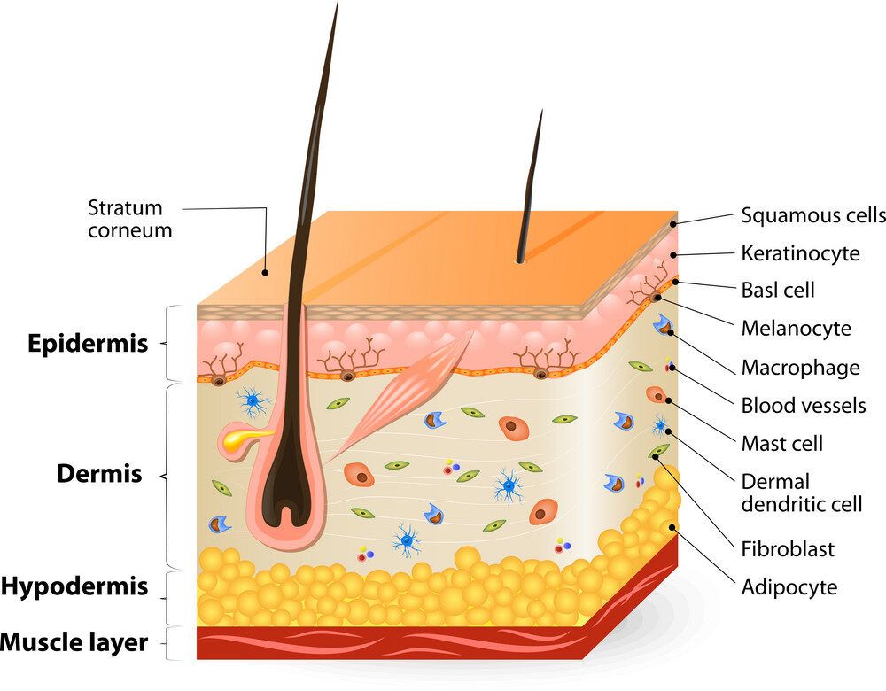
Skin has four identifiable layers
- Epidermis
- Basement Membrane
- Dermis
- Hypodermis
Epidermis:- The epidermis is the outermost layer of the skin and is visible to the eye it provides protection to the body. It is mainly composed of keratinocytes (95% of epidermal cells are keratinocytes). Keratinocytes synthesise keratins which are insoluble proteins. Epidermis is also made up of Melanocytes, Merkel cells and Langerhans cells.
Melanocytes are of Neural crest origin. They produce a protective skin darkening pigment known as Melanin.
Merkel cells are also known as Merkel-Ranvier cells or tactile epithelial cells. They are present very close to the nerve endings and act as mechanoreceptors or transducers for touch.
Langerhans cells are bone marrow derived dendritic cells, they are likely to induce the first
Epidermis is itself divided into five layers the fifth layer is present in some areas of the body. From deep to superficial the layers are as follows:-
- Stratum basale
- Stratum spinosum
- Stratum granulosum
- Stratum lucidum
- Stratum corneum
Stratum Basale :- The stratum basale is the base layer of the epidermis it germinates cells of the epidermis hence also known as stratum germinativum. This is the deepest layer of the epidermis, made up of cuboidal or columnar basal cells.
Function:- Keratinocytes are produced in this layer, as new cells are constantly being formed old cells are pushed out and away from stratum basale.
Stratum Spinosum:- The prickle layer, present between the stratum basale and stratum granulosum. It consists of eight to ten layers of keratinocytes and desmosomes ( helps in cell-to-cell adhesion).
Function:- this layer also consists of Langerhans cells which are tissue resident dendritic cells of the skin. These cells function as macrophages and engulf foreign particles, damaged cells and bacterias in this layer.
Stratum Granulosum:- also known as granular layer. Keratinocytes migrated from the stratum spinosum layer are present here; they are more flatter and irregular in shape, and they have keratohyalin granules which contribute to the keratin content of the cornified cells.
Function:- keratinocytes in this layer create permeability barrier to water and facilitate cell adhesion.
Stratum Lucidum:- it is a thin, translucent smooth layer of the epidermis, present between Stratum Granulosum and Stratum Corneum. This layer is found in thick skin of palms of hands and soles of feet. It is composed of dead, and flattened keratinocytes.
Function:- the keratinocytes in this layer are surrounded by a clear protein called Eleiden, rich in lipids which gives this layer transparent appearance and creates a barrier to water.
Stratum Corneum:- also known as Brick wall and also Horny layer as it is tougher than other layers, it provides protection to the layers beneath it. It is composed of 15 to 20 layers of cells, which are shed and replaced periodically.
Function:- the stratum corneum is composed of keratin a protein which makes up the hair, nails, horns, calluses, claws and outer layer of the skin of vertebrates.
It prevents water loss or absorption of water.
Maintains body temperature at a healthy level.
It also prevents toxins and bacteria from entering the skin.
Basement Membrane:- The basement membrane acts as an anchor for the epidermis but also allows free movement of nutrients between dermis and epidermis. It is composed of well defined two layers, the basal lamina and the underlying layer of reticular connective tissue. These two layers are together known as Basement Membrane. It provides tissue specific functions throughout the body.
Basement membrane is divided into four zones:-
- The Hemidesmosome: stud like structures found in keratinocytes that act as anchoring plaques that connect the epidermis and dermis.
- The Lamina Lucida: found between epithelium and underlying connective tissue like epidermis and dermis.
- The Lamina Densa: along with lamina lucida known as Basal lamina. Lies just beneath the lamina lucida and above the dermis. Provides structural strength to the basement membrane as it contains collagen and other proteins.
- The Sub-lamina Densa: lies below the lamina densa contains anchoring filaments, collagen and elastic fibres.
Dermis:- lies between the epidermis and the hypodermis, also known as corium layer. It consist of dense irregular connective tissue which provides strength to the skin and cushions the body to stress and strain. The dermis is connected to the epidermis through a basement membrane.
Structural components of the dermis are elastic fibres, collagen, mechanoreceptors which are responsible for sense of touch, Thermoreceptors are also present which provide sense of heat, sebaceous glands, hair follicles, sweat glands, lymphatic vessels, nerve and blood vessels are present.
Dermis is divided into two layers:-
- Papillary Dermis
- Reticular Dermis
Papillary Dermis:– it is the uppermost layer of dermis and contains loose areolar connective tissue. It is called papillary because of finger-like projections called papillae that extend towards the epidermis and contain a network of Meissner’s corpuscles.
Reticular Dermis:- present below the papillary dermis, it is vascularized and innervated. It is made up of irregular connective tissue, collagen, elastic fibres. Sweat glands, roots of hairs, sebaceous glands are structures present here. It also consist of langer’s lines also known as lines of cleavage which serve a surgical purpose.


Leave a Reply
You must be logged in to post a comment.